Dental and Medical Simulation Workshop!

Dental and Medical Education Simulation Workshop with Rich Fidler PhD, Dr. Mary Sturgeon DMD, Dr. Melissa Lin DDS and the VAMC SF Medical Simulation Team

Date, Time, Location: Wednesday, March 13th, 2019; 7:30 – 8:30 pm at Terra Linda HS in San Rafael, Room 207

Back by popular demand! Join us for an introduction to dentistry, including the educational requirements and job opportunities. Drs. Melissa Lin, DDS and Mary Sturgeon, DMD will tell you how it’s done. There will be a hands-on break out session including mirror skills. Brace yourselves for an unfolding case presentation– a 70 year old man with symptomatic chin swelling, large anterior mandibular multilocular radiolucency of the anterior mandible! (Spoiler alert – the biopsy shows ameloblastoma.) You can learn about initial evaluation by a general dentist, and evaluation in a specialty consultation by an oral surgeon. Another hands-on break out session will feature a punch biopsy on chicken hearts.  Finally, there will be an oral pathologist evaluation by Jill White, DDS who will present with slides. Join us and learn!.

Links:

VA Advanced Fellowship Program in Clinical Simulation

Facebook event

Natalie Ciaccio, PhD

Title: “The Pharmacy of Genes: Drug Development for Genetic Diseases” with Natalie Ciaccio Ph.D. of Biomarin

Date, Time, Location: Wednesday, October 3rd, 2018; 7:30 – 8:30 pm at Terra Linda HS in San Rafael, Room 207

NEW Watch Dr. Ciaccio’s presentation in VR below!

Bio: Dr. Ciaccio is a Sr. Scientist working in Formulation Development at BioMarin Pharmaceutical, Inc. in Novato, Ca. Prior to joining BioMarin, Dr. Ciaccio completed her Postdoctoral training in the Department of Bioengineering and Therapeutic Sciences at the University of California San Francisco, where she explored the application of drug delivery technologies for sustained release of biologics. Previously, Dr. Ciaccio obtained her PhD in Pharmaceutical Chemistry from the University of Kansas, where she investigated mechanisms of protein degradation and aggregation. She obtained her BS in Pharmacy from Purdue University and spent three years working in Quality Control at Eli Lilly and Co., supporting insulin manufacturing prior to attending graduate school.

Watch Dr. Ciaccio’s presentation in Virtual Reality (VR) below! Use your keyboard arrows or click and drag to visit the Marin Science Seminar classroom.

The Pharmacy Of Genes from Marin Science Seminar on Vimeo.   Video by MSS intern Satvik Namburu

Links:

Join us and learn! – Back to the Marin Science Seminar calendar

Interview with Art Wallace, MD PhD on Big Data and Medical Innovation

By Angel Zhou, Branson School


Mobile technologies, sensors, genome sequencing, and advances in analytic software now make it possible to capture vast amounts of information that could transform medicine. The question is: can Big Data make health care better?

In the upcoming Marin Science Seminar, “Big Data and Medical Innovation,” Dr. Art Wallace, Chief of Anesthesia Service at the San Francisco VA Medical Center and a Professor of Anesthesiology and Perioperative Care at UCSF Medical Center, will discuss applications of Big Data in medicine and how Big Data has changed epidemiology, quality improvement, and drug discovery. Read the following interview to learn more about Dr. 

Wallace’s thoughts on Big Data and its impact on medical innovation.
Art Wallace, MD PhD

What is Big Data and what is its significance to medicine?  What makes Big Data different from other data that people work with in the healthcare industry?
Big Data is data that is acquired for other purposes that can be analyzed to understand processes, people, and systems. Big Data includes many things: cell phone records, super market purchase card records, credit card records, medical records, internet search terms, medication usage, hospital admissions, social security records, etc. This data can be used for epidemiology to identify associations between factors and outcomes.
Big Data gives additional power to identify factors associated with rare outcomes. I can now easily do a study in 1 million people using data collected for administrative purposes. Doing a study in 1 million patients used to be enormously expensive, now it just requires computer programming and epidemiologic analysis. Before Big Data, the cost of collecting data was prohibitive, so many studies could not be done. With Big Data, there is little to no cost of collecting the data, making the analysis the entire cost for large studies. The profoundly lower costs with Big Data techniques make studies that were previously impossible, possible at minimal cost.
How does Big Data impact professionals in the medical field? Can Big Data be used to improve healthcare?
We have identified factors associated with adverse outcomes, identified medication practices that are associated with increased mortality, identified medications that can reduce morbidity and mortality, and we have identified possible therapies for diseases that have no current therapy. We can reduce morbidity, mortality, cost, and assist in the development of new therapies.
  
Big Data can be used to reduce morbidity, mortality, cost, and improve efficiency. Big Data can be used to ask questions that are morally, politically, technically, socially, ethically, or legally impossible to answer with randomized trials. Big Data is being used to improve quality of life while lowering costs.
Describe how Big Data is reshaping the drug industry?
Big Data can be used to identify medications that reduce or increase risks. Post marketing testing can identify medications that have significant associated morbidity and mortality. For example, we identified a drug that increased mortality risk 5 fold (increased from 3 to 15% with drug use). This use of Big Data led to a medication being taken off the market. It had been used in Europe for 30 years, in the U.S. for 10 years, and it increased the risk of death from 3 to 15%. Big Data was used to identify a very serious risk to patients and led to the medication being taken off the market.
How will Big Data accelerate innovation in medicine?
Big Data will be used to identify new uses of medications. It will identify risk factors for morbidity and mortality. It will lead to further randomized trials.
What are the benefits and dangers of providing Big Data online as the “ever expanding cloud of information” becomes more accessible?
It is easy to identify people from their digital detritus. It is easy to identify very personal things about people from their data trails. Factors such as financial status, interests, sexual orientation, political beliefs, religious beliefs, health status, pre-existing medical conditions, drug and alcohol use, pregnancy status, and proclivities can all be assessed via Big Data. Big Data can be used to manipulate, track, and market to people. At the same time, Big Data can identify very serious risks to patients’ health. Scientific method is an approach; Big Data is a tool. Both can be used for good or bad purposes. Big Data is simply a new and extremely powerful scientific tool.   

Join us Wednesday, February 11th, 2015 to learn more about “Big Data and Medical Innovation” with Dr. Art Wallace from 7:30 – 8:30 PM Terra Linda High School, San Rafael in Room 207.

Saving Our Ocean Friends: An Interview with Dr. Claire Simeone of the Marine Mammal Center

by MSS Intern Isobel Wright, Tamalpais High School

From sea lions with cancer to stranded motherless seal pups, Dr. Claire Simeone knows just what to do. Dr. Simeone works as a Conservation Medicine Veterinarian at The Marine Mammal Center in Sausalito, California and at the National Marine Fisheries Service in Washington, DC. In addition to tending to sick animals, she travels the world to attend Unusual Mortality Events, international training programs, and works on the Marine Mammal Health Map. Dr. Simeone attended the University of Maryland College Park to receive her BSc in Physiology and Neurobiology, and graduated from veterinary school at Virginia Tech. Read the following interview to learn more about life at the Marine Mammal Center and working with animals. 
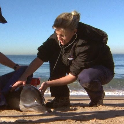
Claire Simeone, DVM
            Could you walk me through your typical day at The Marine Mammal Center?
One of the best things about working at The Marine Mammal Center is that every day is different. Some days, you’re caring for harbor seal pups that have been separated from their mother. Another day, you’re treating California sea lions with cancer. You might be medicating elephant seals that are dying of lungworms. Some days, you’re treating all of those animals, plus caring for the two hundred additional animals that are ALSO onsite. 
As a veterinarian, I usually start my day walking around the pens to check in on all of the animals on-site, and then our team starts procedures, which include blood draws, x-rays, and surgeries. If animals die, we perform post-mortem exams to determine why they died. At the same time, our volunteer crews (more than 1,000 committed people!) are preparing fish, feeding the animals, and cleaning their pens. Our night volunteer crews take care of the animals into the night, and the veterinarians and technicians are on-call 24 hours a day to make sure all of the animals receive the care they need.
What are the best and worst parts of your job?
There are so many best parts of my job. First, I’m lucky to be able to travel around the world to care for marine mammals and learn more about them. Second, I really feel that I’m making a difference with the work I’m doing – whether it’s saving a seal pup or training the next generation of marine mammal veterinarians. Third, I’m constantly learning new things – about marine mammals, their habitats, and what affects their health. 
Because I do work with animals, a difficult part of the job can be seeing animals that are suffering, often because of things humans do – but it helps to know that we are doing everything we can to bring that animal back to health.
What does it feel like to rescue an animal?
Imagine getting a call from someone who was on vacation, and saw a California sea lion that had fishing line around his neck. First, you feel focused – you take down the description of the animal from the citizen, check your maps, and plan out your strategy. Your rescue volunteers have confirmed that this animal is one you’ve been watching for months, and he’s asleep on the beach. You load up the truck, and make the drive to meet your team. You feel hopeful – he’s still snoring away. Holding your breath, you sneak up slowly, and then with a leap you throw the net over his head. He roars as he jumps up and finds himself trapped. With swift action your team boards him into a carrier, and as stealthily as you came, you load him into the truck. You feel elated as you watch him resting calmly on the way home. 
After a quick procedure to remove the line, it’s clear his wound will heal on its own, and he’s ready to go back to the ocean. After driving him back to the beach, you open the carrier, and he strides out into the waves and dives under the break. You feel proud that you’ve saved this animal’s life, and returned him to his ocean home. 
What’s the most common injury/disease you see in marine mammals? How can we prevent this?
Unfortunately, we commonly see injuries that are due to something called human interaction – entangled in fishing line, nets, or plastic packing straps; ingesting pieces of plastic; struck by a boat; or gunshot. In 1972 the Marine Mammal Protection Act was passed, making it illegal to harass or harm a marine mammal. However, many marine mammals are still harmed in passive ways from our trash or discarded items. You can prevent these entanglements by properly disposing of plastics, and helping to keep beaches clean by picking up any trash you see. Just a few weeks ago the annual International Coastal Cleanup Day brought 54,000 volunteers to California’s coasts. They removed over 680,000 pounds of trash in one day!
What level of education and experience do you need to obtain a job like yours?
As a veterinarian, I have a bachelor’s degree, as well as a DVM – Doctor of Veterinary Medicine. However, there are many ways that you can be involved with marine mammals or ocean conservation – through a Master’s or PhD, if you’re more science-focused, or you can have a completely unrelated career, and get your fill through volunteering at a facility like TMMC. We even have a Youth Crew volunteer program for teenagers 15-18 years old (learn more at http://www.marinemammalcenter.org/Get-Involved/volunteer/youth-crew ). As far as experiences go, I would recommend doing as much as you can to get a variety of experiences, which will help you decide what is really right for you. I’ve worked with dogs and cats, horses and cattle, birds and seals, and each experience set me up for the next step in my career. 
What have you learned from working with these animals?
I’ve learned that in order to conserve energy while diving, some seals can lower their heart rate to 10 beats per minute, and right before they surface, their body speeds the rate back up to 120. I’ve learned that a sea otter, if left alone, will unscrew all of the screws on a drain – that were placed with an electric drill! – with its bare paws. And I’ve learned that a harbor seal, blind from cataracts, can find fish by sensing the water movement with its vibrissae (whiskers). Each one of our patients has given me great stories with which to share the knowledge I’ve learned. 
What is an Unusual Mortality Event? What is it like to attend one? Tell me about the most recent one you attended? 
If a group of marine mammals are sick, they may strand on the beach near one another. Unusual Mortality Events (UMEs) are declared when the number of sick or dying animals is larger than expected in that area or time frame. A panel of experts is then called to lead a response to care for the animals, and to try to figure out why they are dying. A recent UME was close to home – in 2013, more than 1500 starving California sea lion pups washed up on southern California beaches. Thanks to the UME response team, it was determined that the reason the pups were starving was because the fish their moms were feeding on had moved farther offshore – meaning they had to go farther to forage. This caused moms to either lack the milk they needed to nurse them, or abandon their pups completely. Caring for hundreds of sea lion pups at a time is exhausting – most need to eat 3-4 times a day, and they may need treatment for vomiting, diarrhea, or pneumonia. It was thanks to hard-working rehabilitation centers, like TMMC, all along the California coast, that we were able to save so many pups. 
What is the Marine Mammal Health Map? How do you contribute to it?
Think about all of the animals we’ve talked about – starving sea lions, entangled elephant seals, gunshot animals or animals with cancer. Each one of these animals provides a unique look at what is happening in the ocean at that location. All of the animals that come through TMMC have a record with all of their health information. Similarly, all of the stranding centers across the country have records on all of their animals. However, there is no centralized database to collect these data, or display them for all to see. The Marine Mammal Health Map will be that space – so that biologists, veterinarians, and members of the public will know what’s happening to marine mammals in their area. I’m working with scientists from around the country to develop the Health Map and ensure that all of our marine mammals are represented. You’ll have to come to the talk to learn more!

Watch this video below to see the process of the rescuing, rehabilitation and release of a sea lion…

Join us for “Sick Seals and Seizing Sea Lions: What Marine Mammals Can Tell Us About the Health of Our Oceans” with Claire Simeone DVM of The Marine Mammal Center, Sausalito – Wednesday, October 8th, 2014 at Marin Science Seminar

High Tech Mannequins

by Gillian Parker, Tamalpais HS   
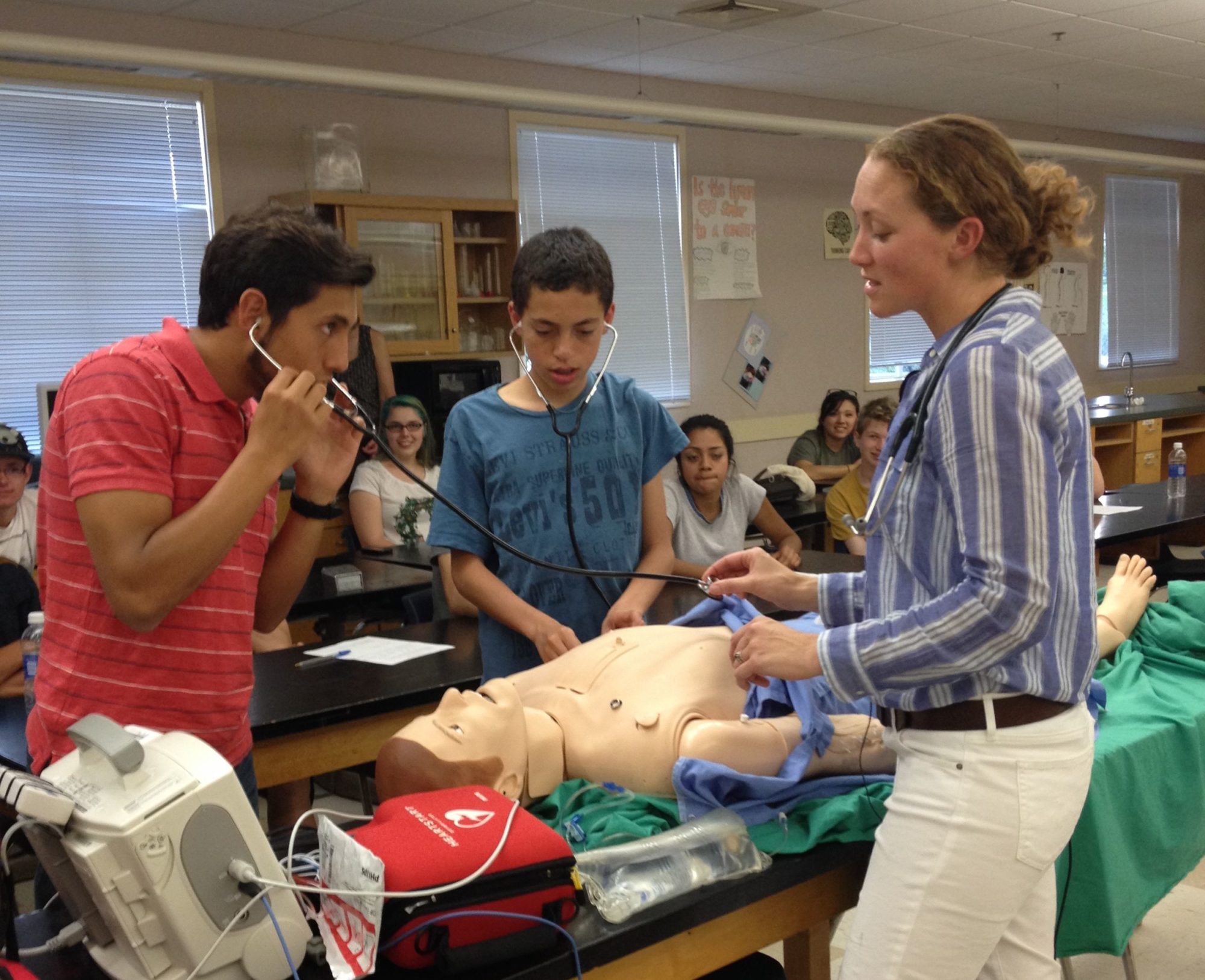
Oftentimes it is nerve-wracking or even dangerous for new medical staff to carry out certain procedures on real patients. At the VA Medical Center in San Francisco, the Simulation Center has high tech mannequins to help train staff in a low-risk environment. These mannequins simulate a normal patient and allow new staff to practice various procedures like chest tube insertion and IV catheter insertion, among others. They can also be hooked up to monitors that are often used to observe patients.

     Abi Fitzgerald practices one day every week in the emergency department at the SFVA as part of her one year fellowship in advanced clinical simulation. She is an RN and achieved her MSN at San Francisco State University. Read the following interview with Abi Fitzgerald to find out more about her experience with the simulators!


1. What is the best part about having the high tech mannequins to practice on?
    
    The manikin’s ability to simulate human functions allows clinicians to practice going through the physical motions assessing patients in both emergent and non-emergent situations, as opposed to verbally walking through the process.  This develops muscle memory and skills for recognizing normal vs. abnormal breath sounds, heart sounds, mental status, neurological functions and more.  

2. How would medical staff be trained without these medical robots?
    They could practice on actors, in which case the abnormal functions such as wheezes or heart murmurs are difficult to simulate.  They would also practice on real patients, which they still currently do, but using a manikin allows them more freedom to perform procedures and other tasks that they wouldn’t necessarily be able to do on a live patient. Working with manikins allows students and clinicians to refine their skills before working with actual patients.
3. What procedures have you performed on the simulators? Could you describe some?
This year the VAMC sim lab acquired a few new simulators that have allowed us (the simulation fellows) and the clinicians who train on them, to become more familiar with a lot of new procedures.  We now have an endovascular trainer which simulates procedures that take place in the cath lab such as non-open heart valve replacements.  This is when the doctors access the heart valve through a long wire and tube that is inserted in the leg and follows the artery all the way up to the aorta and into the heart where they can replace replace a heart valve using fluoroscopic imaging. We also have a new manikin that simulates ultrasounds and displays a three dimensional virtual reality image on a computer screen.   
4. Are there any flaws/negatives to the simulators?
   
The cost of acquiring the simulators and the repair costs can be high, but the quality of training and knowledge gained are very much worth it.  Additionally, ongoing research projects have resulted in the acceptance of grant proposals which has made the acquisition of some of the simulators possible. 
5. What are some things that the simulators can’t fully prepare you for?
  Even though we do our best to make simulated scenarios as real as possible, there are always some elements such as smells and unexpected outcomes that can be difficult but not impossible to simulate well.

Come to the Marin Science Seminar on Wednesday, May 14th, 7:30-8:30 to hear Abi Fitzgerald and Richard Fidler talk about medical education robots at Terra Linda High School in Room 207, 320 Albion Way, San Rafael, CA 94903

The Adorable, Rewarding, and Sometimes Difficult Task of Birthing Babies

 by Claire Watry, Terra Linda HS

A common misconception is that the only job of a midwife is to deliver babies. In reality, the duties of a midwife begin long before the baby is born. A midwife can screen for diseases, prescribe certain 

medications, help the mother stay healthy during pregnancy, monitor the baby’s growth before and after 
birth, provide emotional support, discuss care options and breastfeeding, support the mother and
her child after birth, and provide many more services in addition to the delivering the baby. As a 
midwife, upcoming MSS speaker Sheri Matteo is responsible for the health and wellbeing of a woman
and her baby for the entire length of the pregnancy and beyond. The video below shows the benefits of 
midwifery for women. 


 Matteo explained that midwifery in the U.S. has undergone many changes throughout history.
“Before the early 20th century, most babies in the US were delivered by midwives at home. Then 
medicine, mostly male doctors, brought birth into hospitals and birth became a medical condition. 
Women were put to sleep and babies were delivered with forceps. Moms were encouraged to bottle 
feed. Midwifery pretty much vanished. In the 1970’s, with the rise of feminism and the “natural birth” 
movement, midwifery as a profession started to grow again. Unlike many other industrialized countries
with better childbirth statistics where midwifery care is the norm, midwifery in the US has stayed on 
the fringe and been seen as an “alternative” birth option. In the 15 years that I have been practicing, 
I have seen many midwifery services grow and disband based on whatever economic issue was driving
healthcare at that time. Healthcare in the US is still largely driven by economics and legislation, but 
midwives and other “mid levels” or “allied health professionals” are rising in use and stature. We 
have rigorous education and certification maintenance programs and are fully integrated into healthcare
teams with nursing, case managers and social workers, as well as doctors. The profession is growing in
numbers and strength and our good outcomes are more commonly recognized by clients and medicine
professionals.” According to Time magazine, approximately 8% of babies born in the U.S. – a record 
high – and about 24% of babies in New Mexico were delivered by midwives in 2009. While delivery in 
the U.S. usually takes place in a hospital in the presence of obstetricians, more and more women are 
turning to midwives. 

Read the rest of the interview with Sheri Matteo for a sneak peek preview of her upcoming MSS 
presentation Birthing BabiesWhat it Takes to be a Nurse Midwife”. 


How did you decide to become a midwife?  
I was premed in college and always thought I wanted to be an obstetrician. I was also a singer and performed in HS and college plays. I spent my junior year abroad studying acting in London and when I returned I realized I no longer wanted to go to med school. I received my BA in Theatre Arts at Brown University. I worked as an actress in NYC, off-Broadway and as a member of a theatre company. Working on a play with an older actress, I discovered her daughter was a midwife. I didn’t even know midwifery was a profession at that time! Shortly after, I met a woman who was an obstetrician. I was really inspired hearing about her work. She invited me to “take call” with her one night at the hospital. I saw a birth and I was hooked! Midwifery appealed to me more than medicine because of its emphasis on childbirth as a natural part of life, not a medical condition that needs to be “treated”.


How does one become a midwife? What sort of training or education is necessary?  
There are many different kinds of midwives in the US and several different ways to become one. There are “lay” midwives who receive traditional training as an apprentice of another lay midwife. These midwives are sometimes state licensed and sometimes not. They are not eligible to work in hospitals so they only do home birth.  To be a Certified Nurse Midwife, I had to enroll in nursing school with the plan to continue onto graduate school. I went to Columbia University School of Nursing in NYC. They have a special nursing program for people who already have a baccalaureate. Instead of four years, I earned my BSN (Bachelors of Science in Nursing) in one and a half years. I continued on to complete my Masters in Nursing (MSN) in another one and a half years, for a total of three straight years and two degrees.
What are the responsibilities of a midwife? What is a typical day?  
Certified Nurse Midwives can perform nearly all aspects of women’s health primary care. We screen for diseases, including cervical cancer, breast cancer and STDs. We can prescribe medicine including birth control and antibiotics. We perform prenatal care, making sure moms stay healthy in pregnancy and their babies are growing well.  We can deliver babies in the hospital, in birth centers, or at home. We support women with breastfeeding and take care of them postpartum. We do a lot of client teaching about their health, bodies, and care and treatment options. Though we do not perform surgery like c/sections, we are hands-on surgical assistants. A typical day involves either working at the clinic seeing prenatal patients (between 12-20 in 8 hours) or working in the hospital supporting moms in labor and delivering babies. Because I am the Director of my group of 12 midwives, I also have office work and a lot of meetings to attend. I work on marketing my group’s services, hospital quality and safety and employee satisfaction and performance.


What is the most exciting, rewarding, or difficult aspect of being a midwife?  
Watching new families grow! Helping women be and stay healthy. Meeting all kinds of people from all different cultures. Working with women and empowering them to believe in their natural ability to birth and mother.
What advice do you have for young people aspiring to be in a healthcare profession?  
It is very hard work but also very rewarding. Never forget to treat all people with dignity and respect.


To learn all about midwifery and birthing babies, attend the Marin Science Seminar presentation Birthing Babies: What it Takes to be a Nurse Midwife” with Sheri Matteo, RN, CNM of Prima Medical Foundation, Marin General Hospitalon Wednesday, March 26, 2014, 7:30 – 8:30 pm, Terra Linda High School, San Rafael, Room 207. See the flyer here

For more information go to:
Our Bodies Ourselves Health Resource Center
Dimensions Healthcare System
Student Medics
Time Magazine

Image credits:
https://drewstarr.wordpress.com/pictures/early-american-midwifery/
http://www.sciencemuseum.org.uk/broughttolife/themes/birthanddeath/childbirthandmedicine.aspx
http://healthsciencetechnology.wikispaces.com/Midwife

http://organichomebirth.com/
http://firststepskent.org/welcome-home-baby/welcome-home-baby-book/

~Claire Watry

What Makes a Cancer Cell

by Sandra Ning, Terra Linda HS

Cancer is most commonly treated through radiation, surgery, and chemotherapy.

    While it could be considered cliché to compare cancer cells to supervillains, the similarities are undeniable. Supervillains are cunning, deeply rooted within their far-reaching schemes, and fearsome to the extreme. Cancer cells are just as sly, difficult to remove from the human body and terrifying to the afflicted and their loved ones. It’s not hard to visualize cancer cells as the shady criminal syndicate of the human body; their reach extends to the lungs, bones, tissue and bloodstream, and their tactics are ruthless. Make no mistake—cancer cells have long been antagonists to the scientists fighting for a cure and the patients fighting for their life.
    But when it comes down to the science of it, cancer cells differ from many classic villains in that they aren’t innately evil. Rather, cancer cells and their dangerous properties originate from chance mutations during the division of normal cells. Mutations explain a lot of strange phenomena, from unexpected eye colors to increased resistance to diseases. These unexpected changes in gene sequences can be harmless, or even beneficial. However, they have an equal chance of damaging DNA, mutating it in such a way that the cell distorts into fast-splicing cancer cells.
     Usually, mitosis—the process in which a cell divides—takes precautions against such mutations. “Checkpoints” during a cell’s growth period scan for identity-changing DNA mishaps, ensuring things are running as expected. If something is wrong, the cell will stop growing; if the damage to the DNA can’t be repaired, the cell will kill itself in a process called apoptosis. Through such self-sacrificing vigilance, cells that are mutated beyond repair never get the chance to multiply into a runaway number of damaged cells. But sometimes cell mutations go undetected, due to the sheer number of cells within the human body, with its trillions of constantly dividing cells, each with their own double-helix sequences and enzyme and lysosomes. In such a rush, a handful of mutations can slip by even the strict quality standards cells hold to themselves. Many of these mutations go undetected because they’re harmless to the identity of that cell—but some aren’t so benign.

Normal and cancer cell division. Most damaged cells die through apoptosis.

     When a cell with damaged DNA successfully slips by and divides, it creates the first two in a series of cells that will rapidly divide and spread incorrect DNA, beginning the first rapidfire stages of cancer. The speed of growth and division of cancer cells is unmatched, and unyielding; a cancer cell’s daunting ability to keep multiplying without ever dying, as normal cells do, is often referred to as ‘immortality’. This trait is due to two substances within the cell in particular: telomere and telomerase.
      Telomere is a repeating DNA sequence that essentially acts as a cap for the chromosome it’s on. The sequence acts as a buffer between valuable DNA sequences within the chromosome and the often messy process of dividing a cell. Without the telomere, the ends of the chromosome would lose important base pairs much like a rope fraying at the ends. The more a cell divides, the more telomere is lost in protecting the chromosome. Once all of the telomere is gone, the chromosome reaches “critical length” and no longer replicates. When this happens, the cell doesn’t divide and dies through apoptosis. The erosion of telomere thus measures the age of a cell, with long telomere sequences indicating young cells and short sequences indicating old ones.

The repeating TTGGGG sequence is telomere; the enzyme and RNA template belong to telomerase, which rebuilds worn-down telomere.

     To restore and keep the cycle of cells replicating in our body, telomerase is needed to extend the eroding telomeres. Telomerase is an enzyme made of proteins and RNA. As an enzyme, telomerase enables certain reactions that couldn’t happen without it—in this case, rebuilding and elongating telomeres to a longer sequence again. Telomerase is sparingly used in somatic, or body, cells, which comprise most of the human body. As a result, humans age without much interference from telomerase.
     While telomerase is rarely active in normal body cells, the enzyme becomes ten to twenty times more active in cancer cells. The abundance of telomerase gives cancer cells an endless supply of telomere, and with it, the ability to multiply indefinitely.
    In addition to ‘immortality,’ cancer cells have several additional unique properties that explain why finding a cure is proving so difficult. In addition to fast replication, cancer cells don’t undergo apoptosis easily; high levels of survivin, a protein, inhibits the usual method of cell death. Cancer cells need neither the physical space nor the same amount of nourishing chemicals, known as growth factors, that normal cells need. Instead, they pile freely on top of each other, and remain undeterred by a diet on growth factors. The clusters cancer cells often find themselves in form the lumps within the breasts and testes that doctors and outreach campaigns warn about. Despite their ability to clump, cancer cells have unfortunately high mobility, too. While normal cells anchor themselves onto neighboring cells, cancer cells can break away and travel through the body, infecting other organs. Their ability to invade and infect other areas is made possible through the ability to break through the lamina. The lamina is a noncellular shield that protects the tissues, organs and surfaces within the human body, deflecting normal cells with ease. Cancer cells don’t have the same limitation, and spread to different organs with relative ease.
     With its unique properties, cancer remains frustratingly difficult to cure. Treating cancer needs to somehow overcome the mobility and speed of replication cancer cells exhibit. Current treatments for cancer actually do better than that—the chemotherapy method of treatment uses the cancer cells’ speedy multiplication against it. Chemotherapy sends chemicals throughout the body that kill fast-replicating cells. Cancer cells are efficiently targeted and wiped out through this method, being some of the fasted replicating cells in the body.
     However, chemotherapy has serious faults in its accuracy; by targeting fast-replicating cells, chemotherapy hits hair and blood cells particularly hard. A broad swath of helpful cells get caught in the crossfire between chemotherapy and the cancer cells it’s meant to target. As a treatment for cancer, chemotherapy can cause hair loss, amongst other more painful side-effects.

Chemotherapy affects the fast-growing hair cells as well, which is why cancer patients’ hair often falls out.

     Other treatments are available, when cancer cells are concentrated in specific parts of the body. Radiation focuses on a single area, maybe one organ, to destroy cancer cells. When cancer cells are concentrated in a single area, forming a tumor, surgery can excise the infected part. Sometimes, a mixture of the three treatments are required to treat a patient.
     There is still no way to accurately target and eradicate cancer cells without collateral damage. For that reason, and for the growing number people with breast cancer, leukemia, and other forms of cancer, research for better treatment and ultimately a cure is incredibly important. Cancer is internal, deadly in its silent machinations and intimidating with its arsenal of lethal properties. It’s up to the bright minds and generous hearts of every scientist, doctor, donor and activist to combat, quite literally, the enemy within.

Interested in cancer cells and what scientists are doing to treat it? Come see Dr. Brad A. Stohr present “Why do Cancer Cells Grow Forever and Can we Stop Them?” Dr. Stohr will be presenting this Wednesday, April 17th, at the Marin Science Seminar. The Marin Science Seminar takes place during 7:30 to 8:30 p.m., in rm. 207 of Terra Linda High School. Come check out the Marin Science Seminar on our website and Facebook!

Sources:

Sandra Ning

Interview With Dr.Susan Fisher

by Julia McKeag, Terra Linda HS

Susan Fisher, Ph.D. is the Director of Translational Research in Perinatal Biology and Medicine at UCSF. She is also a Professor in the Departments of Oral Biology, Pharmaceutical Chemistry, and Anatomy and Faculty Director at the Biomolecular Resource Center, UCSF. She is also a member of the UCSF Biomedical Sciences Graduate Program (BMS).

(Figure 1- refer to end of interview) 
What type of experiments does your lab do?
We study the early stages of human development. One of the approaches we use includes deriving human embryonic stem cell lines.
How did you become interested in stem cell research?
Stem cell research is rooted in developmental biology, which I have been interested in for as long as I can remember. I have always been fascinated by how one cell becomes an entire human being.
How do you think stem cell research will benefit humanity?
Eventually we will understand how to cure human diseases using cell-based therapies.
 
(Figure 2)
Do you think Stem cell research will continue in the future despite its surrounding controversy?
Yes. We have learned so much already using stem cell models. This is a very compelling reason for continuing these lines of investigation.
Are animal stem cells similar in structure and function to human stem cells?
There is not a clear-cut answer to this question. We know from comparative analyses that there are similarities and differences. My personal conclusion is that work in both areas is important.
 
(Figure 3)
What is the most interesting thing you’ve discovered about stem cells during your research?
We have developed a new method of deriving human embryonic stem cells that appear to be less differentiated than analogous cells derived by standard methods.
What is an average day as the Director of Translational Research in Prenatal Biology and Medicine at UCSF like? What does this position entail?
I am also head of the UCSF Human Embryonic Stem Cell Program. as Director of Translational Research, I lead programs in which we study placental function in normal pregnancy and in pregnancy complications. My job in the Human Embryonic Stem Cell Program focuses on embryonic rather than placental development. Therefore, between both jobs I get to study the cells that form the placenta and the offspring, which it supports. The work is mesmerizing and extremely rewarding! We get to ask questions about processes that very few people get to study.
Figure 1: Human Embryonic Stem Cells- in a recent medical case, Doctors at Glasgow’s Southern General Hospital grew stem cells into neural stem cells, then injected them into a stroke patient’s brain

Figure 2: Cluster of Human Embryonic Stem Cells
Figure 3: Humans, Animals, and Plants have clusters of stem cells that sustain growth and replace damaged tissues.  


Join Dr. Fisher and Marin Science Seminar this Wednesday to learn more!
Wednesday, March 28th
From 7:30 to 8:30pm
Terra Linda High School
Room 207
Julia McKeag

Interview with Edward Hsiao MD PhD of UCSF

by Julia Moore, Drake HS

How did you become interested in musculoskeletal disorders?
I’ve always been interested in the skeleton. Although we typically think of bones as being solid and unchanging, they undergo a variety of very significant events throughout our lifetime, including growing and repairing after injury. In addition, bones are central to us as a living organism. They provide structure to our bodies, protect soft or vital organs, allow us to move efficiently, and provides bone marrow space for blood formation. We now know that many medically important diseases including osteoporosis, atherosclerosis, and heterotopic bone ossification are all a result of problems affecting normal bone formation.
How are we currently treating different types of musculoskeletal disorders?
Since we don’t  understand how many musculoskeletal disorders develop, our ability to prevent them is pretty limited. Treatments for established disease are also very rudimentary and mostly symptomatic. For example, many inherited diseases of the bone can only be treated by surgery to remove the affected bone. In some cases, we can use metal implants or joint replacement, but these have a relatively short lifespan. Even common diseases, such as osteoporosis or arthritis, have only limited medical treatments.
How do you do your research?
My research is driven by a desire to understand how hormones and genetics control human skeletal growth. Since getting samples of diseased tissues from patients is often difficult, I use a variety of model systems to study skeletal disease. This includes mouse models where I can control hormone signals, and human stem cells created from patients with genetic skeletal diseases (human induced pluripotent stem cells). Together, these models are helping us understand what causes disease and how we can develop new treatments.
What are artificial hormones and how are they advancing research and treatment?
Nature uses hormones as a way to communicate between different parts of the body. One major class of hormone molecules is called G-protein coupled receptors (GPCRs). Since there are over 500 GPCRs in the human genome, figuring out what each individual receptor does is a huge challenge. Our strategy uses a synthetic receptor that only responds to a synthetic drug. This system acts like an artificial hormone – if we add the drug, we can turn the system on; if we take away the drug, we can turn it off. This system allows us to “mimic” a normal hormone system and control that pathway using our drug. This model has proven useful for studying hormone signaling in complex organ systems, including cardiac disease, the brain, and now bone.
What do you think is the future of treatment and prevention of musculoskeletal disorders?
I think that developing robust prevention strategies is important. We also need to develop better combinations of surgical and medical management that have fewer side effects. Much of this can be gained by a better understanding of what happens in normal growth and how those mechanisms go wrong in disease. Finally, I believe that human stem cells provide a valuable new tool in this effort by allowing us to study lab-derived human tissues directly. These stem cells are already providing insights into some rare and dramatic bone diseases. We hope to be able to extend our findings to more common disorders.

Edward Hsiao will be speaking at Terra Linda High School in Room 207 on
Wednesday February 29th at 7:30-8:30pm

Written by: Julia Moore 

Invention in Medicine: How Medical Devices get Invented and Go to Market


with Art Wallace M.D. Ph.D.
December 10, 2008, Room 207

Dr. Wallace started out in experimental surgery and radiology studying imaging of the heart using CT scanners. He has worked on a number of devices that originally were built for experimental studies that evolved into clinically useful devices including a cardiac output monitor, the off pump CABG, off pump aneurysm surgery, electronic sedation, and a selective coronary vasodialtor. Dr. Wallace will explain his experiences with the inventive process using examples from both device design and drug development. There will be a brief discussion of the importance of intellectual property, patents, venture capital, FDA approval, and business development in completing the invention process.